

You watch as a medical team turns a CT scan into a plan, then into a custom implant that restores both function and appearance. Advanced imaging, AI-assisted modeling, and 3D-printed implants let surgeons rebuild a shattered jaw faster and more precisely than traditional methods.
She once faced surgeries to stabilize life-threatening damage; now she walks out with a tailored solution that reduces operating time and improves alignment with her teeth. The article will explain how surgeons, engineers, and labs work together, the tools they use, and why this matters for complex facial reconstruction.

How a Devastating Injury Changed a Teen’s Life
Mya Buie suffered a gunshot that destroyed much of her lower face and jaw, forcing rapid decisions about life-saving care and reconstructive options. A Des Moines trauma surgeon and a surgical team used advanced imaging and 3D planning to stabilize her and map a path for later reconstruction.
Mya Buie’s Story and the Traumatic Shooting
Mya Buie, a teenager from Des Moines, survived a shooting that caused catastrophic facial and jaw damage. The projectile fractured her mandible, displaced bone segments, and caused soft-tissue loss that left her unable to eat and speak normally.
Family members rushed her to a local hospital where trauma teams assessed airway, bleeding, and neurologic status. They documented open fractures and contamination, which increased infection risk and complicated reconstructive timing.
The injury upended Mya’s daily life. She required feeding through a tube for weeks, missed school, and faced months of surgeries and therapy. Emotional and social recovery became as important as rebuilding bone and tissue.
Initial Emergency Treatment and Stabilization
Emergency providers prioritized airway security and hemorrhage control. A Des Moines trauma surgeon performed rapid assessments using CT scans to identify fracture patterns and foreign bodies. Imaging helped plan immediate steps and later reconstructive surgery.
Surgeons cleaned the wound, removed nonviable tissue, and placed drains to reduce infection risk. They used temporary maxillomandibular fixation when possible to maintain alignment and protect soft tissues for future reconstruction.
Antibiotics, tetanus prophylaxis, and pain management started immediately. The team coordinated with plastic and oral-maxillofacial specialists to schedule staged reconstruction, including possible 3D-printed guides and implants that later informed the complex rebuilding of Mya’s face.
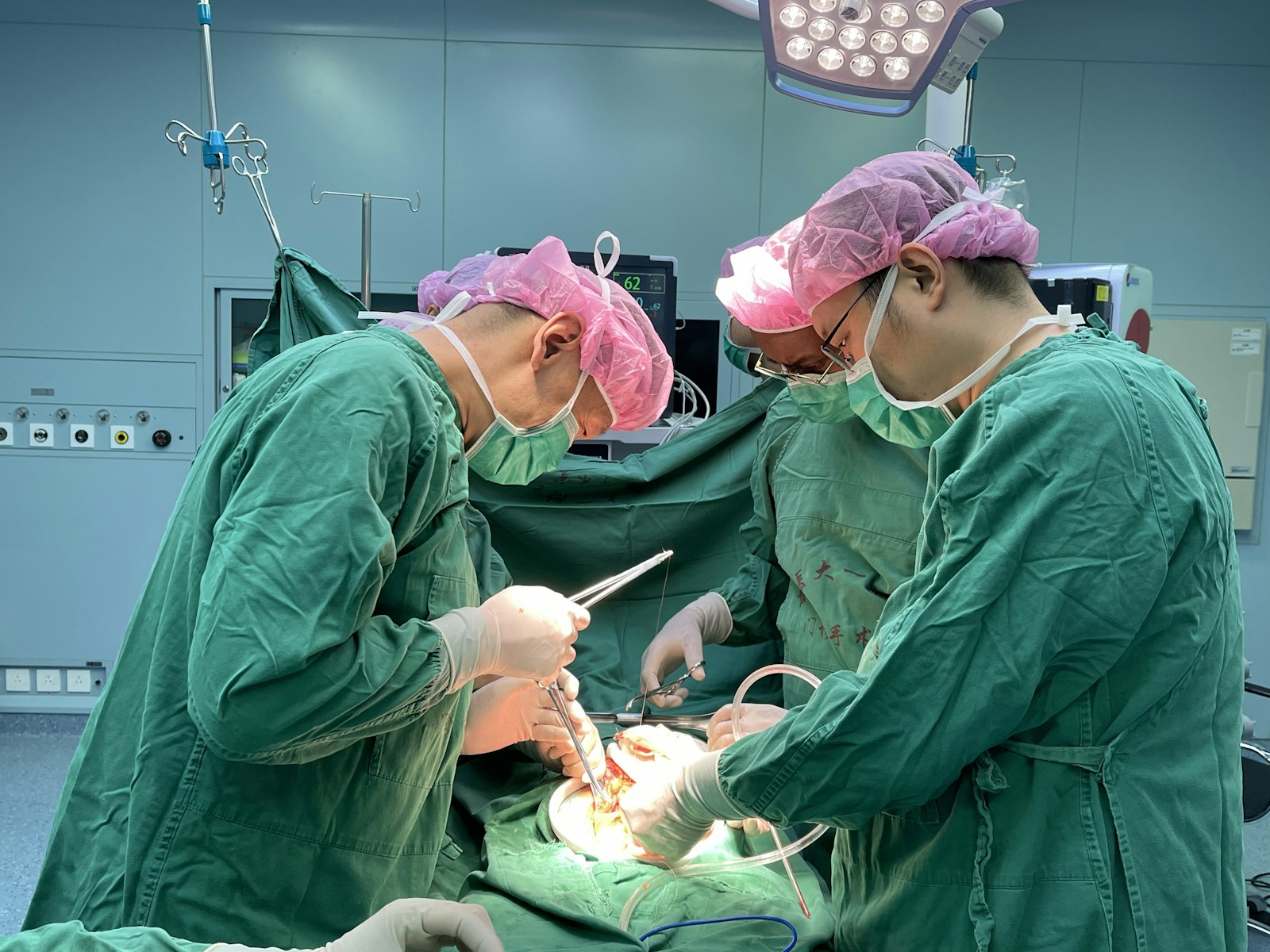
The Role of Surgeons and Specialists
A coordinated team manages complex facial reconstruction, balancing immediate wound care, structural rebuilding, and long-term function. They select technologies and sequence procedures to restore bone, soft tissue, airway, vision, and sensation.
Dr. Simon Wright’s Approach
Dr. Simon Wright emphasizes precise preoperative planning using 3D CT imaging and virtual surgical planning to map bone fractures and design patient-specific implants. He combines intraoperative navigation with custom cutting guides so osteotomies and implant placement match the digital plan within millimeters.
Wright prioritizes staged surgery: first stabilizing airway and soft tissues, then reconstructing bony scaffolding with vascularized bone grafts or custom titanium plates. He integrates microsurgical free flaps when soft tissue volume and blood supply are insufficient, ensuring reliable perfusion for healing.
He uses intraoperative imaging to verify alignment and employs multidisciplinary rounds to adjust timing of revisions, scar management, and sensory nerve repair. Rehabilitation and close follow-up for feeding, speech, and psychosocial needs are part of his protocol.
Head and Neck Specialists in Facial Reconstruction
Head and neck specialists coordinate airway management, nerve repair, and oncologic-grade flap techniques when trauma destroys local tissue. They perform microsurgery to transfer vascularized bone and soft tissue, restoring contour and enabling dental rehabilitation.
These specialists focus on functional outcomes: reestablishing mastication, swallowing, and facial expression through nerve grafts or dynamic muscle transfers. They work with prosthodontists for dental implants and with radiologists for repeated imaging to track bone union and implant position.
Teams usually include anesthesiologists versed in difficult airways, speech therapists, and pain management experts. Collaboration with plastic surgeons, ENT surgeons, and oral-maxillofacial surgeons ensures the technical plan addresses both structural reconstruction and the patient’s long-term quality of life.
Advanced Technology in Facial Reconstruction
Surgeons now use precise imaging and digital models to map bone, soft tissue, and nerves before the first incision. These tools let the team plan cuts, choose grafts, and fabricate patient-specific implants with higher accuracy.
Artificial Intelligence and CT Scans
AI algorithms process high-resolution CT scans to segment bone, cartilage, and critical vascular structures automatically. This reduces manual tracing time from hours to minutes and highlights fracture lines and bone defects with consistent thresholds.
The models from AI-guided segmentation feed into measurements for implant sizing and flap planning. Surgeons can simulate osteotomies, predict postoperative alignment, and assess airway patency before anesthesia.
AI also flags areas at risk for poor perfusion based on prior imaging and helps prioritize tissue reconstruction steps. In clinical reports, combining AI with CT has improved planning efficiency and objectivity in complex craniofacial reconstructions.
3D Virtual Models in Surgery Planning
3D virtual models recreate the patient’s face and skull from imaging and let teams rehearse the operation in software or on a screen. They use mirrored anatomy from the uninjured side to design bone contours, guiding osteosynthesis and soft-tissue draping.
Those virtual files often convert to STL format for 3D printing of anatomical models, cutting guides, and custom implants. Printed models give tactile feedback in the OR and help shape autologous bone grafts or prefabricated alloplasts to exact dimensions.
Teams coordinate between radiology, engineering, and the operating room using the same digital files, shortening operative time and improving fit of implants in reconstructive surgery.
Precision Surgery: Custom Implants and Micro-Surgery
Surgeons combine patient-specific implant design with fine micro-surgical techniques to restore jaw form, bite alignment, and the ability to chew and speak. Rapid digital planning and millimeter-level surgical control speed care and improve functional outcomes.
Designing and Printing Customized Jawbone Plates
Surgeons begin with a high-resolution CT scan to map the fractured mandible and neighboring tooth roots. Engineers use that scan to create a 3D model that matches the patient’s native jaw contours and dental occlusion.
The team selects titanium for strength and biocompatibility, or PEEK for lower weight, then prints the plate or cutting guides with additive manufacturing. Printed cutting guides let surgeons harvest and shape bone grafts — often from the fibula — to match angles precisely during reconstruction.
Speed matters: digital planning and off-site production can cut lead time from weeks to days, allowing earlier fixation and reducing infection risk. Custom plates also improve alignment for future dental implants and lower the need for intraoperative bending.
Benefits of Cutting-Edge Micro-Surgery Techniques
Micro-surgery gives surgeons the dexterity to reattach tiny nerves and blood vessels, which preserves sensation and supports bone graft survival. They use operating microscopes and 1–3 mm sutures to reconnect vessels that sustain free fibula or scapular flaps used in facial reconstruction.
Precise vessel work reduces flap failure rates and limits scar tissue that can impede later dental implant placement. Microvascular techniques also allow single-stage reconstruction: bone, soft tissue, and skin are transferred and inset in one operation, shortening total recovery time.
When combined with custom implants and guided osteotomies, micro-surgery helps restore jaw continuity, enable future dental implants, and improve long-term function and appearance.
Global Innovations and Leading Centers
Several hospitals and research centers combined clinical skill, robotics, 3D printing, and custom implant design to repair complex facial trauma. Teams coordinated imaging, rapid prototype manufacture, and surgical rehearsal to cut operative time and improve fit for patient-specific implants.
Case Studies from Mafraq Hospital and Morriston Hospital
At Mafraq Hospital, Dr. Ali Al Suwaidi led a team that used CT-based surgical planning and patient-specific titanium plates to reconstruct zygomatic and orbital defects. The team prioritized fast in-hospital turnaround for imaging-to-manufacture so implants arrived within days, reducing ischemia time and repeat procedures. They integrated intraoperative navigation to verify implant position against the preoperative plan.
Morriston Hospital applied a multidisciplinary pathway for severe facial trauma under surgeons including Adrian Sugar. They combined virtual surgical planning with mandibular reconstruction techniques and soft-tissue restoration. Morriston emphasized staged reconstruction—first restoring skeletal support with custom guides, then refining aesthetics—while tracking outcomes to shorten recovery and limit revision surgeries.
Role of Cardiff Metropolitan University and PDR
Cardiff Metropolitan University collaborated with the National Centre for Product Design and Development Research (PDR) to translate clinician designs into manufacturable implants. The PDR’s rapid-prototyping labs convert DICOM images into CAD models, then iterate implants for surgeon feedback. That loop lets clinicians such as Dr. John Devine sign off on fit and screw trajectories before production.
The partnership also supports surgical simulation and hands-on workshops so teams rehearse complex approaches. PDR’s expertise in biomaterials and printing tolerances ensures implants meet mechanical and anatomical needs, while Cardiff Met focuses on training and human factors to make the devices practical in real operating-room workflows.
Facial Reconstruction for Complex Cases
Reconstruction combines bone repair, soft-tissue coverage, and functional restoration. Surgeons balance immediate lifesaving steps with long-term plans for chewing, speech, breathing, and appearance.
Treatment for Trauma and Maxillofacial Cancer
Surgeons begin by stabilizing the airway, controlling bleeding, and fixing fractures with plates or external fixation. For bone loss, patient-specific 3D plates and virtual surgical planning guide precise osteotomies and reconstruction; these tools shorten operative time and improve fit.
Soft-tissue defects require local flaps, free tissue transfer, or perforator flaps chosen for volume, vascular supply, and donor-site morbidity. When dental function matters, immediate dental implant placement into fibula or iliac crest grafts can restore occlusion sooner.
When the defect follows tumor resection, oncologic margins dictate how much tissue is removed; reconstructive choices must permit postoperative radiation. Surgeons coordinate timing of reconstruction relative to chemo‑radiation to reduce complications and preserve implant survival.
Teamwork Among Maxillofacial Cancer Surgeons
Complex reconstructions rely on multidisciplinary teams: head and neck surgeons, maxillofacial cancer surgeons, plastic microsurgeons, prosthodontists, and speech therapists. Each member contributes a specific plan: the oncologic surgeon outlines resection, the reconstructive surgeon designs flap and bony reconstruction, and the prosthodontist plans dental rehabilitation.
Intraoperative collaboration uses shared virtual planning files, cutting guides, and navigation to align oncologic goals with aesthetic and functional outcomes. Postoperative meetings track wound healing, implant integration, and radiation timing.
Good team communication shortens hospital stays, reduces reoperation rates, and improves patient function—especially eating, speaking, and facial symmetry—by aligning expectations and sequencing treatments precisely.
More from Vinyl and Velvet:
Leave a Reply